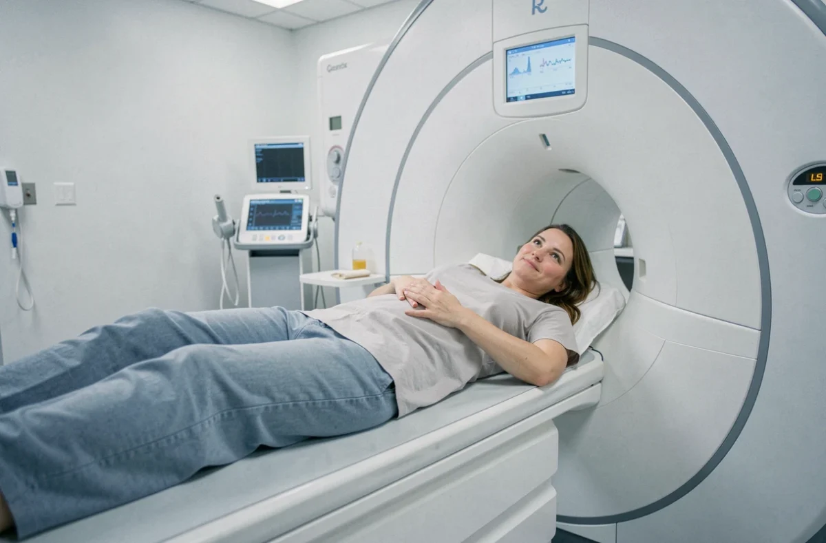
Between five and ten percent of patients experience marked claustrophobia when undergoing an MRI. For these individuals, the examination can become a trial: forced immobility, intense noise, confined space. The open MRI, often touted as the miracle solution, deserves a more nuanced examination than what most online feedback suggests.
Magnetic field and image quality: what the open MRI sacrifices
The open MRI uses a C-shaped or flat magnet that clears the sides of the patient. The visual and psychological comfort is real. However, the magnetic field is often weaker than that of a closed MRI, which reduces the signal-to-noise ratio and the resolution of the obtained images.
This technical limitation is not trivial. For certain fine brain explorations, dynamic liver assessments, or cardiac evaluations, the loss of resolution can compromise the reliability of the diagnosis. Therefore, the type of machine should be validated with the radiologist based on the medical question posed, not solely on the patient’s anxiety level.
Field reports diverge on this point: some radiologists believe that the quality is sufficient for most common indications (lumbar spine, knee, shoulder), while others reserve the open MRI for cases where the examination would simply be impossible otherwise. Before requesting an open machine, it remains relevant to gather feedback on open MRIs for claustrophobics to understand the distinction between perceived comfort and diagnostic relevance.
Wide bore MRI, feet first: alternatives before the open MRI
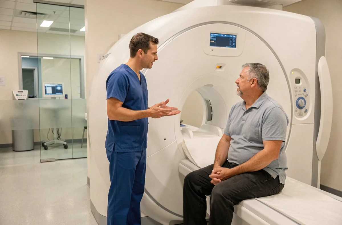
Many patients confuse open MRI with wide bore MRI. The wide bore remains a tube, but with a more generous diameter and reduced depth compared to older models. This difference is enough for moderate anxiety, but not always for intense phobia.
Another parameter rarely mentioned in general content: the entry position into the machine. For MRIs of the spine, pelvis, or lower limbs, a feet-first setup can leave the head outside the tunnel or very close to the opening. This practical detail should be inquired about at the radiology center before the appointment.
The logical gradation looks like this:
- Request a feet-first setup if the examination allows it, on either a classic or wide bore machine.
- Consider a wide bore machine if anxiety remains high despite the appropriate positioning.
- Reserve the open MRI for situations where the first two options fail or cause excessive distress.
- Resort to premedication or, as a last resort, sedation if no material configuration is sufficient.
This gradation avoids mobilizing an open machine immediately, as its availability remains limited in France, with sometimes longer appointment wait times.
Patient testimonials: the role of staff is as important as the machine
Recent testimonials highlight a factor that technology alone cannot replace: human interaction before and during the examination. Patients describe successful examinations on classic closed MRIs thanks to a technician who took the time to explain each step, offered headphones, and maintained regular vocal contact.
In contrast, some feedback on open MRIs mentions disappointment related to a rushed welcome or a lack of information about the actual duration of the examination. The patient’s experience depends as much on the care provided as on the type of machine.
Notify the team when making the appointment
Reporting claustrophobia at the time of booking allows the team to adapt the protocol: longer slots, presence of a companion in the room, choice of installation position. Waiting until the day of the examination to discuss it significantly reduces the staff’s flexibility.
Anxiety management techniques during the examination
Several approaches recur in testimonials from patients who successfully completed their examination despite declared phobia:
- Slow abdominal breathing, practiced a few days before and during the examination, to reduce rising anxiety.
- Keeping eyes closed as soon as lying on the table, even before entering the tunnel, to avoid visual perception of the confined space.
- Using the MRI-compatible headphones provided by the center, with music or a podcast chosen in advance.
These techniques do not eliminate fear. They lower the physiological activation level enough for the examination to proceed to completion.

Premedication and sedation for MRI: what patients report
When non-drug strategies are insufficient, light anxiolytic premedication may be prescribed by the primary care physician or radiologist. The anxiolytic does not eliminate consciousness but reduces emotional reactivity. The patient remains awake, able to follow instructions for immobility and breathing.
Some testimonials describe an effect sufficient to transform a dreaded examination into a bearable experience. Other patients report an effect that is too mild or side effects (prolonged drowsiness, unpleasant floating sensation). The response varies from person to person, making individual feedback not easily generalizable.
General anesthesia remains a rare last resort, reserved for cases where all other options have failed. It involves heavy hospital logistics (anesthetist, recovery room) and risks inherent to any anesthesia, making it disproportionate for most common MRI indications.
The choice between material configuration, human support, and medication assistance is not binary. A patient can combine a wide bore machine, a feet-first setup, and a light anxiolytic for a satisfactory result. The best strategy is built with the radiologist and care team, based on the prescribed examination and the actual level of phobia.